Abstract
Fundus autofluorescence in exudative age-related macular degeneration
Author(s): Q. Peng*, Y. Dong* and P.Q. ZhaoThe aim of this study was to investigate the characteristics of fundus autofluorescence (FAF) in patients with wet (exudative) age-related macular degeneration (AMD). Color fundus photographs, fundus fluorescein angiograms, indocyanine green angiograms, and FAF images were obtained from 61 patients (72 eyes) with exudative AMD. The FAF results for different patterns of exudative AMD were compared to those revealed by other fundus images. Of the 72 eyes evaluated, which were classified into three patterns based on the results of fundus fluorescein angiography, 68 had abnormal FAF. Forty-six eyes (63.9%) had classic wet AMD with abnormal FAF. Among these, 10 exhibited a slightly decreased FAF with near-normal or background FAF signal at the center of the lesion area; 36 demonstrated not only decreased FAF at the center of the lesion but also an increased FAF signal toward the lesion edge. Sixteen eyes (22.2%) had occult wet AMD, of which 12 exhibited heterogeneous fluorescence at the lesion site; 4 yielded normal FAF images. Ten eyes (13.9%) had a mixed pattern of wet AMD with abnormal FAF. FAF imaging suggested that the areas of blood and exudates decreased; however, fluorescence angiography revealed that lesions with hyperfluorescence had background or slightly increased FAF. These results showed that various patterns of wet AMD exhibit different autofluorescence characteristics. These represent the functional and metabolic features of retinal pigment epithelial cells. Therefore, FAF can be used to monitor disease development and evaluate the severity and prognosis of AMD. The aim of this study was to investigate the characteristics of fundus autofluorescence (FAF) in patients with wet (exudative) age-related macular degeneration (AMD). Color fundus photographs, fundus fluorescein angiograms, indocyanine green angiograms, and FAF images were obtained from 61 patients (72 eyes) with exudative AMD. The FAF results for different patterns of exudative AMD were compared to those revealed by other fundus images. Of the 72 eyes evaluated, which were classified into three patterns based on the results of fundus fluorescein angiography, 68 had abnormal FAF. Forty-six eyes (63.9%) had classic wet AMD with abnormal FAF. Among these, 10 exhibited a slightly decreased FAF with near-normal or background FAF signal at the center of the lesion area; 36 demonstrated not only decreased FAF at the center of the lesion but also an increased FAF signal toward the lesion edge. Sixteen eyes (22.2%) had occult wet AMD, of which 12 exhibited heterogeneous fluorescence at the lesion site; 4 yielded normal FAF images. Ten eyes (13.9%) had a mixed pattern of wet AMD with abnormal FAF. FAF imaging suggested that the areas of blood and exudates decreased; however, fluorescence angiography revealed that lesions with hyperfluorescence had background or slightly increased FAF. These results showed that various patterns of wet AMD exhibit different autofluorescence characteristics. These represent the functional and metabolic features of retinal pigment epithelial cells. Therefore, FAF can be used to monitor disease development and evaluate the severity and prognosis of AMD.
Impact Factor an Index

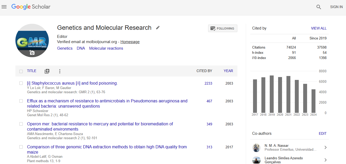
Google scholar citation report
Citations : 74024
Genetics and Molecular Research received 74024 citations as per google scholar report
